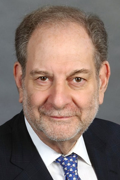
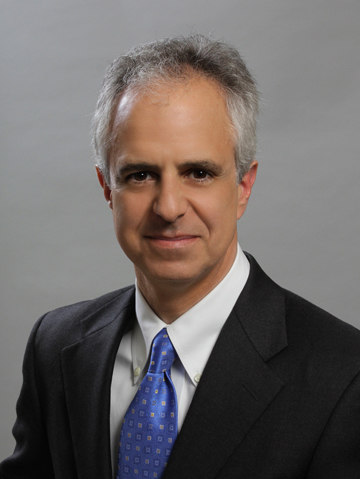
The approval of anti-VEGF therapies more than a decade ago forever changed the retina field. Conditions such as wet age-related macular degeneration (AMD), diabetic macular edema (DME), and retinal vein occlusion (RVO) were no longer blinding; in fact, many patients can achieve good, longterm vision with proper treatment.1,2 But is “good” good enough? Anti-VEGF therapies have limited durability and efficacy, burdening patients with frequent injections for the remainder of their lives.3 Even on maximum treatment, some patients won’t respond. Retina specialists must not become complacent. Several treatments with different mechanisms of action are in the pipeline that may push retinal care into a new era, one that looks beyond anti-VEGF. In the following roundtable discussion, a seasoned panel of retina experts discuss the promise of these therapies and how they can be applied to challenging cases.
— David E. Eichenbaum, MD, Moderator
THE CURRENT TREATMENT LANDSCAPE FOR RETINAL DISORDERS
Q | David E. Eichenbaum, MD: The current standard of care in AMD is anti-VEGF therapy, given every 4 to 8 weeks after three loading doses.4 Dr. Rachitskaya, where is our treatment of AMD succeeding and what are the unmet needs?
Aleksandra Rachitskaya, MD: The current generation of trainees can’t conceive of a world without anti-VEGF for the treatment of neovascular AMD. People used to lose vision and lose it quickly without treatment, and that’s difficult to conceptualize for someone who has never seen it. We’ve made incredible progress, turning neovascular AMD from a blinding condition to a condition we can treat well; blindness from AMD has decreased by 50% since anti-VEGF was introduced.1,2 Solomon et al looked at 12 trials including 5,496 participants with neovascular AMD treated with anti-VEGF. They found that patients who received any of the anti- VEGF agents were more likely to have gained 15 letters or more of visual acuity (VA), lost fewer than 15 letters of VA, and had vision 20/200 or better after 1 year of follow up compared with control arms. We’re able to preserve a lot of vision if treatment is initiated early and maintained.4 That translates into independence and better quality of life.
That said, I see several important unmet needs. First, we don’t have treatments for geographic atrophy (GA). Currently, the only recommend treatment is the use of oral supplements to slow disease progression, based on AREDS2 data.5 There are some promising treatments on the horizon, but we’re not there yet. Second, our treatments for neovascular AMD aren’t durable. The frequency of treatment required is a huge burden on the patient, their caregivers, and the clinic. The Time-and-Motion study found that patients spend an average of 12 hours on every AMD appointment (including travel and post-appointment recovery).3
It’s difficult to reduce that treatment burden with our current agents because we know that frequent injections are critical to optimizing VA. Patients who receive fewer injections lose vision. For example, the SEVEN-UP study showed that in patients who received an average of 6.8 injections of ranibizumab, compared with baseline, half of eyes were stable, whereas one-third declined by 15 letters or more. Of note, patients who received 11 or more injections gained vision.6 Many studies have shown that patients are undertreated, which is one reason why patients in the real world don’t do as well as patients in clinical trials.6,7 A retrospective analysis of nearly 50,000 eyes by Ciulla et al found a direct correlation between the mean letters gained and the mean number of injections during 1 year, with more frequent injections resulting in better vision.8 Holekamp et al found that patients in the real world only receive an average between 4.6 and 6.9 injections, which is well below what’s given on clinical trials.9
Q | Dr. Eichenbaum: Dr. Regillo, what are your aspirations for macular degenerative disease in the coming years?
Carl Regillo, MD: I agree with Dr. Rachitskaya that the main problem with our anti-VEGF agents is they are relatively shortacting. Longer-acting anti-VEGF therapies will help to decrease treatment burden and better maintain the early vision gains over time. Furthermore, although anti-VEGF agents can control exudation and improve vision initially in most patients, recovery of good visual acuity does not occur across the board.10 Improving absolute VA outcomes will also require earlier disease detection, along with finding ways to prevent or minimize atrophy and fibrosis.
CASE 1: Limited Durability Shortens Treat-and-Extend Interval in AMD
Dr. Regillo: Our first case illustrates the issues with limited anti-VEGF durability. This is a 77-year-old woman who presented to me with a 3-week history of visual blur and distortion in her left eye (OS). At presentation, the VA in her left eye was 20/60 and the fundus examination showed signs of AMD with exudation. Her right eye had signs of dry AMD and good VA at 20/25. The fluorescein angiogram (FA) showed some leakage under the central retina OS consistent with type one choroidal neovascularization (CNV). Optical coherence tomography (OCT) OS showed central subretinal fluid (SRF) and findings consistent with CNV, along with some mild overlying neurosensory retinal thickening and edema (Figure 1A). I initiated anti-VEGF therapy with intravitreal aflibercept OS.
Four weeks after the first aflibercept injection, her VA improved by 2 lines to 20/40 with a reduction in the SRF. There was some incidental vitreomacular adhesion, but it was unlikely to be influencing the visual problem at hand. I gave her a second intravitreal injection of aflibercept. Figure 1B shows the left eye 4 weeks after the second aflibercept injection. Her VA is now 20/30 and there is complete resolution of exudation. Are we ready to move to a treat-and-extend paradigm? I think most of us would say “yes” at this point, although some may consider another 1-month treatment interval before starting to extend the follow-up. Also, once we start to extend, will it be by 2 or 4 weeks at a time?
 Figure 1. Case 1: (A) At presentation, the patient’s VA was 20/60 and there was central CNV with SRF on OCT. (B) Four weeks after the first (above) and second (below) aflibercept injections, the patient’s VA improved to 20/20 and there was complete resolution of SRF after the second treatment.
Lejla Vajzovic, MD: I would give three loading doses first. The LUMINOUS study, the first large-scale, global, prospective, observational study, demonstrated that when three loading doses were given, patients had better VA gains (with a 4- or 5-letter) versus if they did not have the loading doses.11 I tend to do standard three injections on a monthly basis. Then after that, I start the treat-and-extend protocol and extend 2 weeks at a time.
David Boyer, MD: I think there’s rationale for both ways. I would extend the patient by 1 week and have her come back at week 5 rather than week 4. You don’t know if you’ve reached the maximum vision improvement at this point. You could argue, certainly, to give her one more injection to ensure the macula is totally dry and see if her VA improves to 20/25 or 20/20. On the other hand, I only go 1 week in a patient like this rather than 2 weeks in the beginning. Then I usually extend by 2 weeks.
Dr. Regillo: I started to extend by 2 weeks. At 6 weeks, her vision was holding well at 20/30 and there were no signs of recurrent exudation on OCT. However, when I attempted to extend to 8 weeks, her VA dropped to 20/50. Figure 2 shows the obvious recurrent fluid 8 weeks after the last aflibercept treatment. There’s been a lot of talk in the last few years about what types of macular fluid (ie, intraretinal [IRF] versus SRF) we can and cannot tolerate; some studies show the presence of subfoveal SRF may be associated with better visual outcomes.12,13 Also, at meetings, it is often said that small amounts of SRF may be acceptable.14 What does the panel think about the fluid shown in Figure 2?
 Figure 2. Case 1: The patient’s VA was 20/50 after extending the aflibercept treatment to 8 weeks. OCT shows recurrent central SRF.
Dr. Rachitskaya: When we look at the post hoc analysis of HARBOR, there is evidence that some SRF could even lead to better visual outcomes; patients with persistent SRF had significantly better VA than patients in which the SRF was completely resolved.15 Some people postulate that if you dry SRF, you can even experience some GA. Zarbin and colleagues compared rigorous treatment for SRF and a more relaxed, tolerant approach.16 They showed there was noninferiority in terms of visual outcomes. But if you look at the number of treatments the patients received, although there was a statistically significant difference, it wasn’t that much of a difference in terms of the number of injections (participants in the relaxed group received a mean of 15.8±5.9 injections and those in the intensive group received a mean of 17±6.5 injections).
In my opinion, not all SRF is created equal. Is it SRF eccentric to the fovea? Is it SRF that’s associated with a history of IRF or is it isolated SRF, like in this case? Is the fluid stable or is it fluctuating? We also need to assess the vision. In this case, the fluid returned and the vision dropped. If it was my patient, I would treat them and cut the interval by 2 weeks. There’s deterioration, especially the drop in vision.
Dr. Vajzovic: I completely agree with Dr. Rachitskaya. The No. 1 factor here is the VA change. To me, this is active disease with loss of vision and presence of new SRF. I would aggressively treat this patient and cut the interval by 2 weeks until vision improved and fluid resolved, and then I would continue that interval for at least three injections. Once stabilized on that interval with improved vision and no IRF or SRF, then I would consider extending interval by 2 weeks again.
Dr. Regillo: I agree that with recurrent fluid associated with a decline in VA, it would be best to reduce the treatment interval to 6 weeks for this patient. That is what I did, and there was full recovery of the visual gain along with complete resolution of the fluid. So now, does the treat-and-extend interval become fixed at every 6 weeks going forward or do you try to reextend at some point? What about switching her to a drug that may last longer? Within the past 18 months, brolucizumab was approved by the US FDA, which, according to the phase 3 HAWK-HARRIER trials, may dry better than aflibercept or last longer to some degree.17,18 Would the use of brolucizumab potentially benefit this patient?
Q | Dr. Eichenbaum: Let’s say we switch her to brolucizumab and get another 2 weeks out of it. I’d argue that the problem will recur a little bit later.
Dr. Boyer, you’re heavily involved in drug development. Are there any drugs in the pipeline that would be appropriate for this patient, if approved?
PIPELINE
Dr. Boyer: There are a number of drugs that could treat a patient like this. Faricimab, a bispecific combination therapy, will likely be available in the future. Phase 3 data from TENAYA and LUCERNE were recently released and showed that nearly 80% of faricimab-treated patients could go 3 months or longer between treatments with superior or identical VA gains as compared with aflibercept.19 Faricimab has two targets: VEGF-A and angiopoietin-2 (Ang-2). Obviously, we know that inhibiting VEGF-A is effective, as that’s how our current treatments function. What’s exciting about faricimab is its ability to inhibit Ang-2, which seems to be an effective approach.
To me, the most exciting of all the treatments for a patient like this is gene therapy. RGX-314 is currently in phase 1/2 trials with 42 patients enrolled. RGX-314 has been well-tolerated so far, with mild, nonserious adverse events such as conjunctival hemorrhage, inflammation, and eye irritation across all five dosing cohorts. Half of patients haven’t needed an anti-VEGF injection in more than 3 years; 67% of patients have been injection-free from between 9 months to 3 years.20 ATMOSPHERE, the first of two planned pivotal trials, is enrolling.
ADVM-022 is being studied in the phase 1 OPTIC trial. Two years after receiving ADVM-022 at the high dose, no one needed retreatment.21 ADVM-022 appears to be well tolerated, with mild to moderate ocular adverse events. Ocular inflammation has occurred, which has responded to steroid drops. ADVM-022 is a single treatment, but it’s not reversible and we don’t know the ramifications of long-term, chronic, low-grade suppression.
KSI-301 from Kodiak Sciences is starting a phase 3 clinical trial. At 1 year, 66% of patients with wet AMD were treatment-free for 6 months or longer; 78% were treatment-free for 4 months or longer.22 The phase 2b/3 DAZZLE trial for KSI-301 has completed enrollment.
The port-delivery system with ranibizumab (PDS) is also promising. 23,24 The PDS had a mean refill time of 15 months in the phase 2 LADDER trial, with 80% of patients not requiring a refill per protocol for the first 6 months.25 The phase 3 ARCHWAY study is currently ongoing, and an open-label extension trial, PORTAL, is planned.
We also have sunitinib, a small molecule multiple receptor tyrosine kinase inhibitor. Sunitinib inhibits VEGF receptors 1-3, blocking all VEGF signals, including VEGF-A, VEGF-B, VEGF-C, and VEGF-D, and placental growth factor. ADAGIO, the phase 1/2a trial, met its primary endpoint of safety and tolerability. Sunitinib reduced injection frequency by 80% with no negative impact on retinal thickness or VA.26 They have had a problem with the drug dissolving and going into the vitreous and anterior chamber, which will need to be addressed in order to move forward.
In summary, there are a number of new drugs on the market that may be more durable and provide better results in the future.
Q | Dr. Eichenbaum: You’ve highlighted fantastic scientific programs with a range of different mechanisms of action. For example, faricimab inhibits Ang-2 and sunitinib is a tyrosine kinase inhibitor. Which of these new mechanisms of action have the most potential?
Dr. Vajzovic: There are several that resonate with me. I’m most excited about gene therapy because of its potential of one-anddone approach. We know AMD cases will explode by 2050; in the United States alone, AMD incidence is expected to rise to nearly 5.5 million by 2050, up from 2 million in 2010.27 A single, durable treatment is the key to managing this dramatic increase in cases. Several gene therapy delivery approaches are currently being studied, and surgical subretinal delivery approach appears to be safe and effective in the RGX-314 trials and the phase 3 ATMOSPHERE study will start soon. I’m also excited about the intravitreal approach in the ADVM-022 OPTIC trial, as this is a convenient, safe and well-established drug delivery method. However, intraocular inflammation appears to be higher with intravitreal injection method and efficacy may be dampened by neutralizing antibodies. Lastly, I am intrigued by a novel suprachoroidal delivery approach in the RGX-314 AAVIATE trial, as it may turn out to be as effective as subretinal delivery method with less risk of intraocular inflammation and convenient and safe approach.
Dr. Eichenbaum: Safety is an important issue. Our current therapies are exquisitely safe. Given that, what is our tolerance for trying something new, albeit with new risks? Brolucizumab had a 4% rate of inflammation in HAWK-HARRIER, 1% of which will result in a severe inflammatory event with vascular occlusion.17,18 That has been a major sticking point for a drug that may have an incremental benefit in efficacy with regard to the anatomy, and durability potentially a little better than what we previously have had.28
Q | Dr. Eichenbaum: How do we balance innovation and safety?
Dr. Vajzovic: That’s an excellent question. The more efficacious the drug is, the more likely we may tolerate safety red flags. Our currently approved drugs have been on the market formore than a decade and are very safe and efficacious. The challenge with brolucizumab is that it has similar to slightly better efficacy to our existing treatments, but there are safety red flags.29 I think whatever drugs are coming down the pipeline need to be at least as safe as the current ones and must more efficacious or at least more durable.
CASE 2: Controlling Disease in a Patient With Idiopathic Polypoidal Choroidal Vasculopathy
Dr. Rachitskaya: Our next case is of a 46-year-old black man with initial 20/20 vision who came in with decreased vision and a subretinal/sub-RPE hemorrhage along the interior arcade, not involving the macula according to the B-scan, as well as vitreous hemorrhage. We suspected he may have idiopathic polypoidal choroidal vasculopathy (IPCV). We gave him several injections, but they didn’t improve his condition, so we moved forward with a vitrectomy. His postoperative OCT showed dominant drusen in both eyes, and old subretinal and sub-RPE hemorrhage inferiorly, but he was dry centrally and no new hemorrhage was seen. I felt the patient was doing great given sparing of the macula, until he came back several months later with new hemorrhage along the inferior arcade and shallow SRF (Figure 3). The patient is still 20/20 and the blood is far from the fovea. How would you approach this patient?
 Figure 3. Case 2: A new hemorrhage in a patient with idiopathic polypoidal choroidal vasculopathy.
Dr. Regillo: This can go very badly, very quickly. Sometimes these polypoidal cases, especially the classic cases with limited polyps in the peripapillary area, can percolate and not behave aggressively but exceptions are not uncommon. It’s just not possible to predict the course at presentation. In my opinion, this one is already demonstrating some aggression, especially with breakthrough vitreous hemorrhage to a degree that necessitated the initial vitrectomy. I take that bleeding to be an ominous sign and so I think this patient needs to be on continuous anti-VEGF therapy to best suppress new bleeding. Right now, we’re lucky that most of the blood and fluid is outside the central macular region, but that could change quickly.
Dr. Eichenbaum: There’s some data indicating our existing anti-VEGF agents have reduced durability in vitrectomized eyes.30 Aspirationally, what would you do? Let’s take gene therapy out of the equation, since that’s relatively early in development. For this patient, would you be happy with the current commerciallyavailable agents, would you want a combination therapy, or would you want to use the PDS?
Dr. Regillo: Those are all excellent options. Faricimab may be approved in the next 2 years, given its positive phase 3 data.19 The ARCHWAY phase 3 trial for the PDS was positive as well, with more than 98% of patients able to go the first 6 months between filling the device without any injection supplements.31 This patient needs continuous, good anti-VEGF effect. We don’t want an injection lapse because that’s when they can have a significant macular bleed. My aspirational treatment would be faricimab, given its dual mechanism of action, but the PDS is also an effective way to deliver a high level of anti-VEGF in a continuous delivery fashion that could help to keep his macula in good shape.
Dr. Rachitskaya: I agree with Dr. Regillo. When I saw the blood, I decided the patient needed treatment. For patients with IPCV, we have a little less guidance than just in standard neovascular AMD, but we still have great trials to turn to like PLANET and EVEREST II. In IPCV, there is a role for both anti-VEGF and photodynamic therapy (PDT).32,33 I started this patient on monthly anti-VEGF injections (initially with bevacizumab and switched to aflibercept rapidly) and gave him PDT. Despite monthly treatments, his vision dropped to 20/500; an extensive hemorrhage continued to develop despite our best effort. This patient was compliant with monthly injections. Figure 4 shows what’s happening on the OCT. He has SRF and sub-RPE fluid that corresponds with the hemorrhage. He’s losing vision and is quite distraught. How would you treat this patient?
 Figure 4. Case 2: Extensive hemorrhage despite monthly anti-VEGF and PDT.
Dr. Boyer: I think aflibercept is the correct drug to use for polypoidal disease. I think you need to evacuate that hemorrhage or the patient may end up with permanent vision loss. I don’t see any advantage of using other anti-VEGF agents, so I would continue to treat with aflibercept. I would also consider pneumatic displacement and if that failed, I would attempt surgical displacement of that blood, which appears to be right in the fovea.
Dr. Vajzovic: I also would have included PDT. I’m definitely concerned about a subretinal hemorrhage here. I would continue with aggressive anti-VEGF treatment at this point.
Dr. Rachitskaya: These are excellent points. I did consider surgery, but I didn’t do it at that time because I was concerned that, given the extensive amount of sub-RPE blood, displacing the subretinal blood might not be sufficient. I discussed the situation with the patient and suggested anti-VEGF injections every 2 weeks. He was eager to try that, but because of insurance issues, I ended up alternating between aflibercept and bevacizumab and it worked. I saw him recently, and his vision is 20/40. The outcome is marvelous, but it also means I’m injecting him every 2 weeks. I’m struggling to envision what I’m going to do for this patient long-term. We can add another round of PDT, but the frequent injections are not sustainable long-term. This is a chronic condition for which this patient will have to maintain treatment to some degree for the rest of his life.
Dr. Eichenbaum: This is the type of eye that needs something more than every 2-week injections. An eye like this could be a good candidate in the future for combination therapy to see if you could modify his disease. This disease has a lot of drive. It could be all VEGF, Tie2/Ang-2, VEGF-C, or VEGF-D. He could even do well with the PDS but may still need booster injections.
I was training in fellowship from 2005 to 2007, and I was fortunate to see retina change dramatically in a short time. Anti-VEGF injections are now part of daily practice, but they used to be a big deal because of endophthalmitis.
Q | Dr. Eichenbaum: Dr. Vajzovic, do you think retina still has the spirit of early adoption and innovation or are we addicted to high-frequency anti-VEGF? Do you think there’s an appetite for something more, whether it is a new delivery system, a new mechanism of action, or a whole new class of treatment, like gene therapy?
Dr. Vajzovic: Yes, I think there is an appetitie for more. I’m very excited about the future options. It’s easy to just continue with intravitreal injections because it’s safe and we’re used to it. But the treatment burden isn’t sustainable. We must address the treatment burden with longer acting agents. I’m excited for VEGF-C and VEGF-D blockade such as OPT-302, combination therapies such as VEGF-A and Ang-2 blockade in faricimab, and potentially having better VA outcomes with the continuous release of a drug such as ranibizumab in the PDS.
Dr. Eichenbaum: I hope we still have an appetite for it, because I agree—there’s much more we can be doing for our patients.
CASE 3: Neovascular AMD With Pigment Epithelial Detachment
Dr. Eichenbaum: Our third case is a 65-year-old woman who came in with a vision change in her left eye. The VA in her left eye is 20/40 and her right eye is 20/25. Her right eye has drusen consistent with dry AMD. Based on Figure 5, what does she have? Is this vascular or is this a serous pigment epithelial detachments (PED) without a neovascular component?
 Figure 5. Case 3: A 65-year-old female with sudden change in central vision.
Dr. Rachitskaya: She obviously has a PED. It’s hard to say if it’s new vascular membrane. She has SRF and maybe some subretinal hyper reflective material. Interestingly, her choroid is not very thin. I would get some additional imaging, either FA or OCT angiography (OCTA). Sometimes it’s hard with the artifacts that you get with PEDs to assess the choroidal neovascular membrane on OCTA.
Dr. Eichenbaum: Her OCTA was not very useful because of the artifacts, as you mentioned. I started treatment with aflibercept. After five aflibercept injections, her VA was 20/50, and I was not happy with the result. I extended treatment. Do you agree with that next step?
Dr. Regillo: It’s questionable whether there’s choroidal neovascularization here, but when in doubt, I think you’re obligated to begin treatment and see what happens. If nothing changes on OCT or vision after five or six treatments, then that will favor this PED not being vascularized. I’ll either start to extend or sometimes even just stop and watch closely. I agree with everything you’ve done. It’s better to err on the side of treatment, especially knowing the safety profile of the current anti-VEGF therapeutics. As long as you have a high level of suspicion that you might be dealing with a masquerade for neovascular AMD, such as a nonvascularized PED, then extending is a logical way to test the waters.
Dr. Eichenbaum: Figure 6 shows the same patient after her sixth aflibercept injection and an extension. Her VA descreased to 20/63. What is my next step?
 Figure 6. Case 3: Six weeks after sixth aflibercept injection.
Dr. Boyer: I don’t think this is neovascularization. I think you’re dealing with a masquerade, and I think this is probably avascular. I would stop treating and observe this patient.
Dr. Eichenbaum: I changed agents and gave the patient one brolucizumab injection to ensure I didn’t miss any opportunity to treat an exudative component. There was no change a week after brolucizumab. I decided to stop treatment. The patient came back several months later and has definitely gotten worse. Her VA is now 20/100 (Figure 7). Would anyone go back to 2-week antiangiogenic treatment in this patient who’s losing vision and becoming a little bit desperate?
 Figure 7. Case 3: Several months after treatment suspension.
Dr. Regillo: I don’t think we could fault anyone for doing either nothing or going back to frequent injections. We’ve all seen those occasional cases of serous PEDs that shrink with frequent or very aggressive anti-VEGF therapy. However, this is probably the natural course of nonvascularized PED with your case, and treatment is unlikely to help.
Dr. Eichenbaum: Do you think combination therapy would help or would it not make a difference? Do you think this eye would respond differently to treatment that targeted Tie2/Ang-2, VEGF-C, or VEGF-D?
Dr. Regillo: If there’s no neovascularization, I doubt anything that’s directed specifically at blocking neovascularization would benefit this eye. That being said, it is possible there are other targets for therapies that could effectively address whatever mechanism is causing such a large collection of fluid under the RPE.
Dr. Rachitskaya: Everyone has excellent points. It’s difficult for physicians to do nothing when a patient’s condition is worsening in front of you. These cases are very frustrating because we don’t know the exact pathogenesis of what’s happening in this patient. I’d have a frank conversation with the patient and ask what they’d like to do. When I see patients like this, I show them their images. It helps with a patient-centered approach and is very important to have shared decision-making. This may be the natural course of her condition. However, if she’s extremely motivated and wants to try more injections, I’d consider it.
Dr. Eichenbaum: I did talk to her, and we decided to reinitiate treatment. Her VA is currently 20/100, and I will be giving her another short series of anti-VEGF to assess for a treatment response, or at least stability. I am doubtful this is a process I can impact with the current armamentarium of drugs, and I’m not hopeful this likely nonneovascular process will respond to something more, but I have elected to try again because she’s getting worse. This patient needs help and is motivated to do something. I don’t want to give up on her.
UNMET NEEDS IN THE TREATMENT OF DIABETIC EYE DISEASE
Dr. Eichenbaum: What is your single most important aspirational wish for diabetic eye disease, specifically DME in the future?
Dr. Vajzovic: When I think of a DME wish list, I think of screening patients early. Telemedicine is the future. We must also continue incorporating OCTA and fundus photography in primary care offices and using them as screening tools to identify disease early and initiate early treatment. There’s mounting evidence from RISE, RIDE, VIVID, and VISTA showing that we can reverse disease the earlier we treat these patients.34,35 Anti-VEGF therapy resulted in visual improvements as early as 7 days postinjection. Improvement in screening is No. 1, followed by earlier treatment.
Dr. Eichenbaum: Earlier treatment, leveraged through community screenings, could certainly save vision. Getting into the primary care office if possible, even getting out to where people live and work has a lot of potential.
Dr. Rachitskaya: Patients with DME are typically working. They have a chronic condition and have had it for a very long time. Adherence to treatment is a huge challenge for these patients, even for patients who have the best intentions.36 It’s difficult for them to maintain good glycemic control. Sometimes they aren’t adhering to our treatments because they are hospitalized or they have complications related to diabetes that affect other organ systems.37 It’s not that they don’t want to come in.
My wish would be a drug that addresses both DME and diabetic retinopathy (DR) because they go hand-in-hand. DR can result in very significant events that can be sight threatening. I would like something with which I could treat a patient and protect them for a longer period of time on both of those fronts, so they don’t get lost to follow-up for a year and then return with 20/400 VA and a horrible tractional retinal detachment.
Dr. Regillo: We need agents that are more effective in drying. We need to get the macular dry faster, and we need more durable agents so we don’t have to inject as frequently.
Dr. Boyer: Diabetes is multifactorial. You may have patients who don’t respond to anti-VEGF at all or patients who are suboptimal responders; instead of gaining 12 letters, they gain 6. This is an area where combination therapies, including steroids, will play a major role. I think the No. 1 thing we need is education of the internist and general practitioner. The HbA1C needs to be below 8%, if not below 7%. Many diabetic patients are never screened, and that’s another area for major improvement.38
Dr. Eichenbaum: Dr. Boyer, you mentioned steroids. That’s really where we do have combination therapy and something other than anti-VEGF that’s commercially available and part of our standard of care armamentarium for many decades now. My aspiration would be to have something with a nonsteroidal, antiinflammatory mechanism of action that would get us the benefits that we see in some steroid-responsive diabetics, but without the steroid-induced cataract and increased intraocular pressure.39
CASE 4: Managing Proliferative Diabetic Retinopathy in a Patient With Poorly Controlled Diabetes
Dr. Vajzovic: Case 4 is a 41-year-old black man with type 1 diabetes. His HbA1c is poorly controlled at 11% and he is on both insulin and an oral hypoglycemic. He has proliferative diabetic retinopathy (PDR) in his left eye, which has very poor vision, and he has undergone several surgeries for a tractional retinal detachment. He was referred to us for the treatment of his right eye, which is 20/20. Figure 8 shows the degree of macular and peripheral retinal ischemia with neovascularization of the disc and periphery in the right eye. I diagnosed him with PDR in his right eye as well. What treatment would you give him?
 Figure 8. Case 4: PDR in patient with poorly controlled diabetes.
Dr. Regillo: I think there are several good options for this patient. One option would be to start with anti-VEGF injections and acheive some good regression quickly, and then follow that with panretinal photocoagulation (PRP) for more permanent, long-lasting neovascular regression effect. You could also start with PRP and forgo the injections knowing there is no DME. Protocol S told us that initial PRP on average will work just as well, and my personal experience is in line with that in eyes with PDR and no DME.40,41 At 5 years, 305 patients in Protocol S received a mean 19.2 injections in the ranibizumab group versus 5.4 injections in the PRP group with comparable visual outcomes.41 If there’s some vitreous hemorrhage, I start with the anti-VEGFs until the hemorrhage has resolved and then I follow it up with PRP. I will occasionally use anti-VEGF alone and not do PRP for PDR, but the patient has to prove to be very reliable with follow-up appointments.42 I wouldn’t take that chance with this patient, given his poor vision in his left eye. I think this patient would really benefit from PRP at some point.
Dr. Vajzovic: I discussed the treatment options with the patient and he was adamant that no PRP be performed, primarily because of the risk of peripheral vision loss, which can impact driving.43 He was very concerned it would limit his capabilities at work. We proceeded with ranibizumab. To Dr. Regillo’s point about reliability, the patient has already demonstrated variable or questionable compliance. The advanced disease, loss of vision in the left eye, and poorly controlled HbA1c tells you he’s not on top of things.
Dr. Rachitskaya: Sometimes when I treat patients like this, I show them their FA and I compare theirs to a normal widefield FA from the Internet. Showing them the dark areas on the image helps them visualize their disease. I’ve also found that involving family members helps them make informed decisions as well.
Dr. Eichenbaum: You need to get buy-in from these patients in the first visit and build that trust or they won’t return. I also like to show patients their widefield FA compared a normal FA and then explain exactly how serious their condition truly is.
Dr. Vajzovic: He did come back for follow-up appointments initially, and I proceeded to give him anti-VEGF injections, and extended them to an 8-week interval. At that point about a year and a half into his treatment, he started missing appointments. It took some convincing, but he eventually agreed to PRP when he returned. He’s done really well since. Remarkably, he had no DME during this process.
Dr. Eichenbaum: That’s remarkable and speaks to the multifactorial process of diabetic eyes. Why does this patient with profound retinal ischemia and some traction never develop DME? Part of the reason is you’re suppressing him periodically with antiangiogenics but he is not a swollen diabetic, he’s a proliferating diabetic. Perhaps in the future, beyond the anti-VEGF era, we’ll be able to tailor treatments to diabetics with certain clinical characteristics or clinical biomarkers, like severe proliferation, severe ischemia, or severe edema.
Dr. Regillo, do you think future therapeutics would give this patient a better result? Would he be a candidate for the PDS?
Dr. Regillo: Yes, he would. It’s being tested now in patients with PDR, both with and without DME. DR often improves to some degree with continuous VEGF blockade, but, more often than not, DME is multifactorial; that’s why you get such a variable or slower response to anti-VEGF alone in some patients. Long-acting anti-VEGF certainly will benefit these eyes. I can’t think of a reason not to inhibit VEGF in a patient with DR. Furthermore, because Ang-2 also likely plays a role in DR and DME pathophysiology, the use of faricimab in this setting makes a lot of sense. At the very least, it appears to be more durable than currently used anti-VEGF agents and with the added Ang-2 blockade mechanism of action, it could provide other benefits in the management of DR and DME.
CASE 5: Addressing Drug Durability Challenges in Branch Retinal Vascular Occlusion Treatment
Dr. Eichenbaum: Dr. Boyer, what advancements in the treatment of branch retinal vascular occlusion (BRVO) would you like to see beyond the anti-VEGF era?
Dr. Boyer: I’d like to see longer acting drugs. We control RVO very well with anti-VEGF but they don’t last long enough; when the anti-VEGF wears off, RVO blows up again and we’re starting over at baseline. Some patients can go 4 or 5 weeks, but they can’t go 6 weeks. It’s also very difficult to get patients off treatment.
Our final case is a 65-year-old male who presented with BRVO about 12 years ago. He has high cholesterol and a history of seizures. I started him on bevacizumab, as we didn’t have many other choices at that time. He received injections monthly, and then every 6 weeks. I added laser treatment to extend the interval, because he couldn’t be extended beyond 6 weeks. After a few treatments, he had a very nice response. His hemorrhages decreased and the veins reduced in size. We held treatment to see if he would improve. About 40% of patients with BRVO will get to 20/40 VA without treatment. I withheld treatment, which was the first mistake I made. Figure 9 shows the OCT at 6-week treatment intervals. I wasn’t acheiving the response I wanted.
 Figure 9. Case 5: Patient with BRVO after 6-week treatment extension.
Nowadays, most clinicians would have switched back to 4-week intervals or maybe another anti-VEGF treatment like ranibizumab or aflibercept. I didn’t have a lot available, so I used a 0.7-mg dexamethasone intravitreal implant (Figure 10). It completely dried him out. I followed him every 6 to 8 weeks, and he did not require another treatment for 3 years. He received one additional 0.7-mg dexamethasone intravitreal implant after 3 years and has remained dry, with 20/25 VA for 10 years.
 Figure 10. Case 5: Patient with BRVO after treatment with a 0.7 mg dexamethasone intravitreal implant.
The point here is that sometimes we continue to treat with anti-VEGF out of habit. But sometimes you have to think out of the box and use another treatment. In this case, his BRVO had some inflammatory component. I have no idea why this patient responded so brilliantly, but I’ve found this in other patiens in whom I couldn’t extend with anti-VEGF and pivoted to the 0.7-mg dexamethasone intravitreal implant, and they’ve done very well.
Dr. Eichenbaum: This is foreshadowing the future. This is a 10-year-old case that shows how going to a different drug class, something beyond anti-VEGF monotherapy, has altered the disease. You may have changed the pathophysiology when you introduced the steroids.
Dr. Boyer: I agree; we need other modes of action. If we could get an anti-inflammatory treatment that doesn’t cause cataracts or glaucoma, we’d use it more frequently. A variety of conditions, including AMD, have an inflammatory component.
LOOKING TO THE FUTURE: NEW THERAPIES WILL ADDRESS TREATMENT SHORTCOMINGS
Dr. Eichenbaum: We’ve done well for our patients with our current standard of care armamentarium, but we’ve all highlighted that there’s a long way to go with efficacy while preserving the excellent safety that we’ve enjoyed for the past 15 years of intravitreal biologic injections.
Q | Dr. Eichenbaum: Dr. Rachitskaya, considering the cases we’ve just discussed, what would you like to see in the future?
Dr. Rachitskaya: Going back to the first question you asked, I think there’s unmet needs in all the conditions we discussed today: AMD, DME, and RVO. The future is bright. I’m excited to see so much work being done to address those gaps in patient care. Time will tell what future treatments are safe, what treatments do well in phase 3 studies, and what treatments really benefit patients in the real world. Patients have reason to hope. We are working extremely hard to find even better approaches than our current treatments.
Dr. Regillo: Our current anti-VEGFs are wonderful drugs; they work well in most of our patients with these common conditions, but there are limitations in durability and efficacy. The conditions have some unique pathophysiologic aspects, and as we head into the future, we’re looking at not only VEGF blockade that is more durable, which is something we certainly want and need, but also introducing other mechanisms of action into the therapeutics, along with better ways to deliver them. That’s going to give us better outcomes in all of these conditions.
Dr. Boyer: I’m looking forward to home OCT and earlier detection of disease. For both DME and AMD, it’s very important we have early detection to get the patient the best vision. I’m looking for longer duration drugs that will allow us to reduce the treatment burden.
We are in exciting times, and it’s extremely fortunate for our patients that we have these additional treatments today. Three or 4 years from now, we’ll have even more treatment options. I’m very excited about future prospects.
Dr. Vajzovic: I echo the desire for early detection, telemedicine, remote diagnosis, and in-home OCT. We also need broader biologics to not just address anti-VEGF, but other modes of mechanism. I’m particularly excited about gene therapy and a one-and-done approach. We have a very bright future ahead of us, and I’m excited to be able to offer our patients more options.
Dr. Eichenbaum: I’d like to thank this group of bright, engaged, and compassionate retina specialists for participating in this discussion. At the end of the day, it’s about helping patients do well while suffering less. That’s where all of us are focused.
Thank you for taking the time to discuss treating retinal disorders in the post anti-VEGF era.
REFERENCES
1. Solomon SD, Lindsley K, Vedula SS, et al. Anti-vascular endothelial growth factor for neovascular age-related macular degeneration. Cochrane Database Syst Rev. 2014;8:Cd005139.
2. Bloch SB, Larsen M, Munch IC. Incidence of legal blindness from age-related macular degeneration in denmark: year 2000 to 2010. Am J Ophthalmol. 2012;153(2):209-213.e202.
3. Prenner JL, Halperin LS, Rycroft C, et al. Disease burden in the treatment of age-related macular degeneration: findings from a Time-and-Motion Study. Am J Ophthalmol. 2015;160(4):725-731.e721.
4. Flaxel CJ, Adelman RA, Bailey ST, et al. Age-related macular degeneration Preferred Practice Pattern. Ophthalmology. 2020;127(1):1-65.
5. Age-Related Eye Disease Study 2 Research G. Lutein + zeaxanthin and omega-3 fatty acids for age-related macular degeneration: the Age-Related Eye Disease Study 2 (AREDS2) randomized clinical trial. JAMA. 2013;309(19):2005-2015.
6. Rofagha S, Bhisitkul RB, Boyer DS, et al. Seven-year outcomes in ranibizumab-treated patients in ANCHOR, MARINA, and HORIZON: a multicenter cohort study (SEVEN-UP). Ophthalmology. 2013;120(11):2292-2299.
7. Maguire MG, Martin DF, Ying GS, et al. Five-year outcomes with anti-vascular endothelial growth factor treatment of neovascular agerelated macular degeneration: the comparison of age-related macular degeneration treatments trials. Ophthalmology. 2016;123(8):1751-1761.
8. Ciulla TA, Hussain RM, Pollack JS, et al. Visual acuity outcomes and anti-vascular endothelial growth factor therapy intensity in neovascular age-related macular degeneration patients: a real-world analysis of 49 485 Eyes. Ophthalmol Retina. 2020;4(1):19-30.
9. Holekamp NM, Liu Y, Yeh WS, et al. Clinical utilization of anti-VEGF agents and disease monitoring in neovascular age-related macular degeneration. Am J Ophthalmol. 2014;157(4):825-833.e821.
10. Krebs I, Glittenberg C, Ansari-Shahrezaei S, et al. Non-responders to treatment with antagonists of vascular endothelial growth factor in age-related macular degeneration. Br J Ophthalmol. 2013;97(11):1443-1446.
11. Hamilton RD, Clemens A, Minnella AM, et al. Real-world effectiveness and safety of ranibizumab for the treatment of myopic choroidal neovascularization: Results from the LUMINOUS study. PLoS One. 2020;15(1):e0227557.
12. Wickremasinghe SS, Janakan V, Sandhu SS, et al. Implication of recurrent or retained fluid on optical coherence tomography for visual acuity during active treatment of neovascular age-related macular degeneration with a treat and extend protocol. Retina. 2016;36(7):1331-1339.
13. Jang L, Gianniou C, Ambresin A, et al. Refractory subretinal fluid in patients with neovascular age-related macular degeneration treated with intravitreal ranibizumab: visual acuity outcome. Graefes Arch Clin Exp Ophthalmol. 2015;253(8):1211-1216.
14. Guymer RH, Markey CM, McAllister IL, et al. Tolerating subretinal fluid in neovascular age-related macular degeneration treated with ranibizumab using a treat-and-extend regimen: FLUID study 24-month results. Ophthalmology. 2019;126(5):723-734.
15. Sarraf D, London NJ, Khurana RN, et al. Ranibizumab treatment for pigment epithelial detachment secondary to neovascular age-related macular degeneration: post hoc analysis of the HARBOR study. Ophthalmology. 2016;123(10):2213-2224.
16. Zarbin M. Anti-VEGF resistant subretinal fluid is associated with reduced risk of macular atrophy and better visual acuity: Drug-induced choroidal new vessel homeostasis? . Association for Research in VIsion and Ophthalmology (Virtual). Paper presented on May 6, 2020; 2020.
17. Dugel PU, Koh A, Ogura Y, et al. HAWK and HARRIER: Phase 3, multicenter, randomized, double-masked trials of brolucizumab for neovascular age-related macular degeneration. Ophthalmology. 2020;127(1):72-84.
18. Dugel PU, Singh RP, Koh A, et al. HAWK and HARRIER: ninety-six-week outcomes from the phase 3 trials of brolucizumab for neovascular age-related macular degeneration. Ophthalmology. 2021;128(1):89-99.
19. Roche. New phase III data show Roche’s faricimab is the first investigational injectable eye medicine to extend time between treatments up to four months in two leading causes of vision loss, potentially reducing treatment burden for patients.
20. Regenxbio. Regenxbio announces additional positive interim phase i/iia and long-term follow-up data of RGX-314 for the treatment of wet amd; 2021.
21. Adverum Biotechnologies I. Adverum Biotechnologies announces positive interim data from cohorts 1-4 from OPTIC Phase 1 Trial of ADVM-022 intravitreal gene therapy for wet AMD; 2020.
22. Kodiak Sciences. Kodiak Sciences announces 1-year durability, efficacy and safety data from ongoing phase 1b study of KSI-301 in patients with wet age-related macular degeneration, diabetic macular edema and retinal vein occlusion at the Angiogenesis, Exudation and Degeneration 2021 Annual Meeting.
23. Chen ER, Kaiser PK. Therapeutic potential of the ranibizumab port delivery system in the treatment of AMD: evidence to date. Clin Ophthalmol. 2020;14:1349-1355.
24. Sharma A, Kumar N, Parachuri N, et al. Ranibizumab port delivery system (RPDS): realising long awaited dream of prolonged VEGF suppression. Eye (London, England). 2020;34(3):422-423.
25. Campochiaro PA, Marcus DM, Awh CC, et al. The port delivery system with ranibizumab for neovascular age-related macular degeneration: results from the randomized phase 2 LADDER Clinical Trial. Ophthalmology. Apr 1 2019.
26. Graybug. GB-102 in Retinal Diseases. www.graybug.vision/our-technologies-and-pipeline/#gb102.
27. National Eye Institute. Age-related macular degeneration (amd) data and statistics. https://www.nei.nih.gov/learn-about-eye-health/resources-for-health-educators/eye-health-data-and-statistics/age-related-macular-degeneration-amd-data-and-statistics. Accessed March 16, 2021.
28. Baumal CR, Spaide RF, Vajzovic L, et al. Retinal Vasculitis and Intraocular Inflammation after Intravitreal Injection of Brolucizumab. Ophthalmology. 2020;127(10):1345-1359.
29. Witkin AJ, Hahn P, Murray TG, et al. Occlusive retinal vasculitis following intravitreal brolucizumab. J Vitreoretin Dis. 2020;4(4):269-279.
30. Edington M, Connolly J, Chong NV. Pharmacokinetics of intravitreal anti-VEGF drugs in vitrectomized versus non-vitrectomized eyes. Expert Opin Drug Metab Toxicol. 2017;13(12):1217-1224.
31. Roche. Phase III data show roche’s port delivery system with ranibizumab enabled over 98% of patients to go six months between treatments for neovascular age-related macular degeneration.
32. Lee WK, Iida T, Ogura Y, et al. Efficacy and safety of intravitreal aflibercept for polypoidal choroidal vasculopathy in the PLANET Study: a randomized clinical trial. JAMA Ophthalmol. 2018;136(7):786-793.
33. Koh A, Lai TYY, Takahashi K, et al. Efficacy and safety of ranibizumab with or without verteporfin photodynamic therapy for polypoidal choroidal vasculopathy: a randomized clinical trial. JAMA Ophthalmol. 2017;135(11):1206-1213.
34. Brown DM, Nguyen QD, Marcus DM, et al. Long-term outcomes of ranibizumab therapy for diabetic macular edema: the 36-month results from two phase III trials: RISE and RIDE. Ophthalmology. 2013;120(10):2013-2022.
35. Heier JS, Korobelnik JF, Brown DM, et al. Intravitreal aflibercept for diabetic macular edema: 148-week results from the VISTA and VIVID Studies. Ophthalmology. 2016;123(11):2376-2385.
36. Weiss M, Sim DA, Herold T, et al. Compliance and adherence of patients with diabetic macular edema to intravitreal anti-vascular endothelial growth factor therapy in daily practice. Retina. 2018;38(12):2293-2300.
37. Gao X, Obeid A, Aderman CM, et al. Loss to follow-up after intravitreal anti-vascular endothelial growth factor injections in patients with diabetic macular edema. Ophthalmol Retina. 2019;3(3):230-236.
38. Centers for Disease Control and Prevention. National Diabetes Statistics Report - 2020.
39. Boyer DS, Yoon YH, Belfort R, Jr., et al. Three-year, randomized, sham-controlled trial of dexamethasone intravitreal implant in patients with diabetic macular edema. Ophthalmology. 2014;121(10):1904-1914.
40. Gross JG, Glassman AR, Jampol LM, et al. Panretinal photocoagulation vs intravitreous ranibizumab for proliferative diabetic retinopathy: a randomized clinical trial. JAMA. 2015;314(20):2137-2146.
41. Gross JG, Glassman AR, Liu D, et al. Five-year outcomes of panretinal photocoagulation vs intravitreous ranibizumab for proliferative diabetic retinopathy: a randomized clinical trial. JAMA Ophthalmol. 2018;136(10):1138-1148.
42. Sun JK, Glassman AR, Beaulieu WT, et al. rationale and application of the protocol s anti-vascular endothelial growth factor algorithm for proliferative diabetic retinopathy. Ophthalmology. 2019;126(1):87-95.
43. Subash M, Comyn O, Samy A, et al. The effect of multispot laser panretinal photocoagulation on retinal sensitivity and driving eligibility in patients with diabetic retinopathy. JAMA Ophthalmol. 2016;134(6):666-672.


